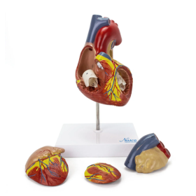

A two times life-size model that describes all the main structures of the human heart in detail. The right part is detachable, to expose the inner chambers and valves. The atrium and wall of the left ventricle can be removed to reveal the atrium, mitral valve, ventricle, and papillary muscles. Made with long-lasting synthetic material. Accompanying an interactive 3D anatomical model with augmented reality is a great tool to encourage learning and support. This platform allows students to engage in comparative analysis of anatomical models as they compare and contrast the structure of individual organs. This initiative also provides a platform for continuing education, providing opportunities for all students to increase their knowledge of anatomy, physiology and pathophysiology.